MARINEMAMMAL RADIOLOGY.COM
Ultrasound
The contents of this page were provided by Dr. Brian Kot.
Applications of ultrasound on cetaceans
The content below summarizes essential experiences and provided information on the applications and limitations of ultrasound in marine mammal medicine.
Cetaceans could be trained to approach the poolside, and coaxed to station themselves in various positions, such as sternal, dorsal, lateral and dorsal recumbency; pinnipeds could be trained to station erect, or to lie down and roll into a supine or semi-supine position on land. Ultrasound examinations using different techniques on various structures could last up to 5 minutes.
Most abdominal, neck and vascular structures are able to be examined by ultrasound for clinical and research purposes. A comprehensive understanding on animal species, physiological status and nutritional conditions should be made for the preparation of and during ultrasound examinations, for efficient and accurate diagnosis. Equipment selection, scanning techniques and normal sonographic anatomy should be well-addressed, along with illustrations of marine mammal pathologies identified using ultrasound. Apart from the traditional B-mode imaging, advanced imaging modes such as 3D/4D volumetric imaging, Color/Power Doppler, Pulsed Wave Doppler could be also applied on various occasions. Limitations included machine inaccessibility, animal uncooperativeness, large body size, thick integument and blubber layers, which prohibited examination of some structures.
Ultrasound is useful in various aspects of marine mammal medicine, and should be incorporated as a tool for artificial insemination and controlled natural breeding, and routine health surveys in a preventive medicine program, as it enables the monitoring of reproductive and maternity cycles, the diagnosis of diseases and the course of therapy.
Thyroid and Adjacent neck structures
For thyroid ultrasound scanning, the transducer should be placed in a transverse orientation at the thoracic inlet, midway between the insertions of the pectoral fins. The transducer should then be moved cranially until the brachiocephalic vein is identified (Figure 1). The transducer should then be moved further cranially until the transverse dimension of the left and right lobes of the thyroid gland is identified. The transducer could then be rotated by 90º to visualize the longitudinal dimension of different portions of thyroid gland. The transducer could also be placed in an oblique manner to visualize the long axis of the thyroid lobes and produce measurements for thyroid volume calculations (Figure 2, for thyroid volume estimation, refer to Kot et al., 2012).
The pretracheal layer of the deep cervical fascia formed a sheath encasing the sternocephalicus and sternohyoideus muscles. These muscles were always well-visualized by ultrasound. Although varying in thickness, the sternocephalicus and sternohyoideus muscles could be recognized as elongated structures at the anterior aspect of the thyroid gland. The sternocephalicus muscle was slightly hyperechoic than the normal thyroid parenchyma, whereas the sternohyoideus muscles appeared as two round structures and was either isoechoic or hypoechoic when compared to the thyroid gland (Figure 3).
The posterior surface of the thyroid gland is related, from front to back, to the trachea. The trachea was identified on ultrasound as a hyperechoic line, corresponding to the anterior wall of the trachea, with posterior acoustic shadowing (Figure 4).
The lateral surface of the thyroid gland was related to the major cervical vessels embedded with the carotid sheath and cricoid cartilage. Major cervical vessels comprised the internal and external carotid arteries (located immediately lateral to thyroid lobes), and internal jugular vein (IJV) (located lateral to the internal and external carotid arteries and more or less spontaneously visible). Cricoid cartilage is located lateral to the carotid arteries and the IJV was identified on ultrasound as an oblique hyperechoic line (Figure 4).
Thyroid lobes appear elliptical or fusiform in the transverse scan plane (Figure 3) and round to oval in longitudinal scan planes (Figures 5 & 6); morphologically, glands comprised 2 lobes joined by an isthmus or a roughly diamond-shaped structure located on the ventral surface of the trachea. Thyroid parenchyma is typically uniform and homogeneous, with echogenic reticulations and well-defined borders (Figure 7). Thyroid glands are hypoechoic or isoechoic relative to the sternocephalicus muscle; echogenicity appears greater in adolescents than in adults. Thyroid gland volume differs between sexes, between sexually mature and immature dolphins, and among age groups and is positively correlated with body length and weight (Kot et al., 2012). Major blood vessels and cervical lymph nodes could be identified (Figures 8 - 10).
The neck lymph nodes could also be visualised by ultrasound. The lymph nodes is isoechoic to hypoechoic when compared to the adjacent sternocephalicus muscle, round or oval in shape, and with an echogenic hilus (Figure 11). The neck lymph nodes are usually found between the sternocephalicus and sternohyoideus muscles, cranial and lateral to the thyroid gland, and around the mediastinum and brachiocephalic veins, at the ventral depth of 40 - 70 mm. The mean long axis measurement of the lymph nodes was 24 mm (ranged from 20 to 35 mm) (Kot et al., 2012).
Lungs
Ultrasound techniques and appearance of the lungs are well-described in Brook et al., 2001. Pleural surface appears as smooth hyperechoic curved line in the near field, with its continuity disrupted by rib shadows. Reverberation artifacts are results from the air-to-tissue interface, which hinders the assessment of deeper lung parenchyma (Figure 12). This typical “normal” pattern will be changed with various pathological conditions such as lung consolidation, abscesses, and fluid accumulation.
Gastric chambers of the stomach complex
Ultrasound techniques and appearance of the gour gastric chambers, named forestomach, fundic chamber, pyloric chamber, and duodenal ampulla, are well-described in Brook et al., 2001. Assessment of contraction rate, wall thickness and specific pattern of gastric rugae could be performed using ultrasound (Figure 13).
Liver
Ultrasound techniques and appearance of the liver lobes are well-described in Brook et al., 2001. Apart from the routine subcostal oblique scan approach, paramedian upper abdominal longitudinal and intercostal scan approach could also be performed to facilitate a full assessment on the porta hepatis, bile ducts, portal veins, and lateral portions of the liver (Figure 14). Ultrasound abnormalities of the liver may consist of diffuse or circumscribed changes in the normal hepatic architecture.
Kidneys and Urinary bladder
Ultrasound techniques and appearance of the kidneys and bladder are well-described in Brook et al., 2001. Bilateral kidneys appear as highly lobulated structures, with multiple echolucent lobules, representing the functional unit renicule (Figure 15). Ultrasound assessment of the bladder and other pelvic structures necessitates a relative full bladder as possible, which facilitates comprehensive assessment of bladder wall anatomy (thickness and focal abnormalities, presence of trabeculation, and diverticula) and bladder capacity in milliliters upon urine collection. The bladder could also a useful anatomical landmark in the abdominal cavity for locating reproductive organs, such as ovaries (Figure 16).
Female reproductive tract
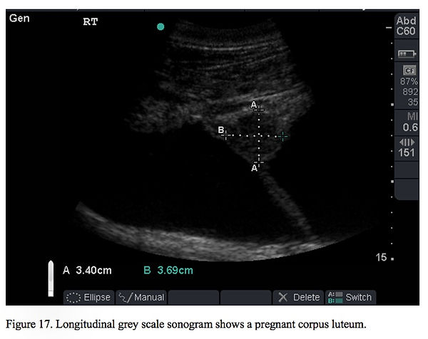
Ultrasound techniques and appearance of the female reproductive tract are well-described in Brook et al., 2001. Ovarian activities, and its corresponding reproductive states could be accurately assessed using ultrasound, including anestrus, estrus (follicular, ovulatory and luteal phase) (Figure 17) and pregnancy. Fetal morphology during the course of gestation could be evaluated (Figure 18) and fetal measurements (biparietal and thoracic diameter) could be performed for estimation of expected delivery date.
Male reproductive tract
Ultrasound techniques and appearance of the male reproductive tract are well-described in Brook et al., 2000 and Brook et al., 2001. Testes, epididymides, vasa deferentia, penis, bulbourethral and bulbocavernosal muscles could be assessed using ultrasound, with the exception of prostate, which is difficult to identify among adjacent muscles. Distinct testicular echopatterns could be used to assess different reproductive status of male dolphins.
References
Brook FM, Kinoshita R, Brown B, Metreweli C. 2000. Ultrasonographic imaging of the testis and epididymis of the bottlenose dolphin, Tursiops truncatus aduncas. Journal of Reproduction and Fertility 119: 233-240.
Brook FM, Van Bonn W, Jensen E. 2001. ‘Ultrasonography’, in Dierauf, L.A. and Guilland, F.M.D. (eds.) CRC handbook of marine mammal medicine: Health, disease, and rehabilitation. Boca Raton, FL: CRC Press, pp. 593–620.
Kot BCW, Ying MTC, Brook FM, Kinoshita RE. 2012. Evaluation of 2-D and 3-D ultrasound in the assessment of the thyroid gland of the Indo-Pacific bottlenose dolphin, Tursiops aduncus. Journal of Zoo and Wildlife Medicine 43(1):33-49.
Kot BCW, Ying MTC, Brook FM, Kinoshita RE. Cheng SCH. 2012. Sonographic imaging of the thyroid gland and adjacent neck structures of the Indo-Pacific bottlenose dolphin, Tursiops aduncus. American Journal of Veterinary Research 73(11):1696-1706.